Keratoconus
If your health fund covers cross linking (Medicare item number 42652) then you can have your procedure done while you are lightly sedated or sleeping at the South Brisbane Day Hospital with NO GAP for the anaesthetist.
The cornea is the clear window at the front of the eye. It is a normally round cornea in a healthy eye. Keratoconus is a degenerative disease of the cornea where it slowly changes shape from being round to developing a cone shape. This irregular, cone-shaped deformation causes astigmatism which causes distorted vision and blurs your vision. The cornea's structure is supported by collagen fibres, which help maintain its shape and integrity. Normally astigmatism can be treated with glasses, however, in keratoconus the astigmatism is too irregular for glasses to fix so special keratoconus contact lenses are required. Keratoconus affects up to 1 in 86 people depending on the racial group. It generally appears in the late teens to early twenties and tends to stabilise by age 40.
Vigorous eye rubbing is a known risk factor for keratoconus and should be avoided.
Traditionally 1 in 4 people with keratoconus needed a corneal transplant. Whilst this is an effective treatment, corneal transplants can reject and fail. Thankfully with modern treatment, newly diagnosed patients will only very rarely need a corneal transplant as we can now halt the progression of keratoconus and even remove the corneal irregularities with laser surgery.
Introduction to Keratoconus
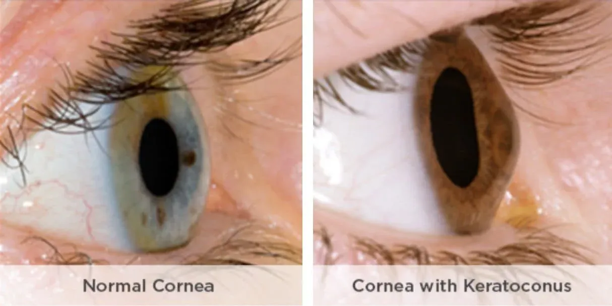
Keratoconus is a progressive eye disorder that affects the shape of the cornea, the clear front surface of the eye. Instead of maintaining its normally round shape, the cornea gradually thins and bulges outward into a cone-like shape. This irregular shape of the cornea leads to distorted and blurry vision, making everyday tasks such as reading or driving more difficult. Keratoconus often begins to show symptoms in the late teens or early twenties and can worsen over time if left untreated. Treatment options for keratoconus range from prescription glasses and soft contact lenses in the early stages, to more advanced interventions like corneal transplant surgery in severe cases. Consulting a keratoconus specialist is essential for accurate diagnosis and to determine the most effective treatment plan for your individual needs. Learn more about the difference between keratoconus and keratoglobus.
Understanding Keratoconus
Keratoconus is a condition that specifically impacts the corneal tissue, causing it to weaken and change shape. The cornea plays a crucial role in focusing light onto the retina, and any alteration in its structure can significantly affect vision. Excessive eye rubbing is a known risk factor for keratoconus, as it can further damage the delicate corneal tissue and accelerate the progression of the disease. Genetics also play a part, with individuals who have a family member affected by keratoconus being at higher risk themselves. Diagnosing keratoconus requires a comprehensive eye exam, which allows your eye care professional to assess the health of your cornea and recommend the most suitable treatment to preserve your vision.
Signs and Symptoms of Keratoconus
The signs and symptoms of keratoconus can differ from person to person, but there are some common indicators to watch for. Many people first notice distorted and blurry vision that cannot be corrected with regular glasses. Increased light sensitivity, frequent eye strain, and discomfort—especially when trying to wear contact lenses—are also typical. In the early stages, symptoms may be subtle, but as the condition progresses, vision problems often become more pronounced, sometimes including double vision or rapidly changing prescriptions. A keratoconus specialist can use advanced diagnostic tools such as corneal topography to map the surface of the cornea, confirm the diagnosis, and discuss the most appropriate treatment options to address your specific needs.
Treatment options for Keratoconus
1. STOP RUBBING YOUR EYES
Many patients with keratoconus have a long history of rubbing their eyes due to chronic allergy. This rubbing, especially vigorous rubbing, can severely worsen keratoconus and should be strictly avoided. It is imperative that you stop rubbing your eyes if you have keratoconus. Topical anti-histamine drops such as Patanol or Zaditen may be helpful in relieving the chronic irritation and itch.
2. CONTACT LENSES
Most patients with keratoconus require specially fitted contact lenses to have good vision. However, some patients may not tolerate contact lenses and may require alternative treatment options. These contact lenses should provide excellent vision but do not treat the actual disease. Generally, specially trained contact lens optometrists are required to fit theses lenses. These optometrists have the specialised equipment and expertise to fit complex contact lenses for keratoconus.
3. COLLAGEN CROSS-LINKING
Patients with worsening keratoconus can have their progression halted with this procedure, known as corneal cross linking or corneal cross-linking. Corneal cross-linking works by applying riboflavin drops to the cornea, which are then activated by ultraviolet light. This reaction increases the number of cross links between collagen fibres, creating strong, permanent bonds that substantially strengthen the cornea and stop the progression of the disease. Corneal cross-linking is an advanced procedure for strengthening the cornea and is effective in halting keratoconus progression.
The procedure can be performed as an outpatient procedure if you do not have private health insurance. This means you can maximise your medicare rebates with the medicare safety net. If you have health insurance then Dr Cronin performs the procedure in a hospital to minimise the cost of corneal collagen cross linking.
Dr Cronin uses the best collagen cross linking machine available. The revolutionary eye tracking technology in the Mosaic KXL2 topography guided collagen cross linking machine means that more energy is delivered to the steeper parts of your cornea. This personalised and customised treatment is individual for each person. It delivers better vision and better outcomes to more patients. Dr Cronin and his colleagues at QEI are the only surgeons in Australia with this amazing cross linking technology.
See a video of the procedure here
4. LASER CORNEAL RESURFACING AND COLLAGEN CROSS-LINKING (“ATHEN’S PROTOCOL”)
This involves using an excimer laser to “resurface” the affected cornea and remove the irregular astigmatism that causes the reduced vision in keratoconus. This is called a phototherapeutic keratectomy (PTK) and it leaves the cornea with a nice regular shape. Immediately after the laser resurfacing has occurred, the cornea undergoes collagen cross-linking, which increases the number of cross links within the corneal structure, enhancing its strength and stability. So, can you have laser eye surgery if you have keratoconus? Yes! But not in all cases and you will need an individual assessment with Dr Cronin to assess your suitability. See a video of the procedure here.
5. CORNEAL ALLOGENIC INTRASTROMAL RING SEGMENTS (“CAIRS”)
In this procedure, a femtosecond laser is used to create a ring-shaped tunnel in your cornea. A small, similar shaped ring of donor corneal tissue is threaded into the tunnel in your cornea. This ring of corneal tissue improves the strength, rigidity and overall shape of your cornea. CAIRS is often combined with collagen cross linking to enhance the strengthening effect. CAIRS has many of the advantages of both Kerarings and other types of corneal transplants without many of the downsides. CAIRS offers tailored solutions for patients with keratoconus, providing personalized treatment options based on individual needs. QEI was the first clinic in Australia to offer CAIRS as a treatment and Dr Cronin regularly hosts surgeons from around the world to teach this technique. He even runs a website that is used by surgeons globally to plan their CAIRS procedures - www.cairsplan.com. Watch a video of the CAIRS procedure here.
6. BOWMAN’S MEMBRANE TRANSPLANTATION
Bowman’s layer is a tough but clear layer in the cornea. In a normal cornea it is approximately 10 microns thick but it is often weakened and degraded in keratoconus. Bowman’s membrane transplantation is considered for patients with progressive keratoconus who require additional corneal strengthening. In a Bowman’s layer transplant this 10 micron layer is delicately peeled from a donor cornea. A femtosecond laser is used to create a pocket in your cornea. The donor Bowman’s layer is then inserted into this pocket. This strengthens and stiffens your cornea.
7. PLASTIC CORNEAL SEGMENTS (KERARINGS)
Plastic corneal segments, such as Kerarings, are one method for treating keratoconus by reshaping the cornea. These small implants are inserted into your cornea to pull the keratoconic cornea back into a better shape, much like “CAIRS” as discussed above. CAIRS has completely replaced Kerarings as a procedure for keratoconus as it is safer and more reliable.
8. CORNEAL TRANSPLANTATION
Corneal transplantation is typically considered when glasses or contact lenses and other treatments are no longer effective in managing keratoconus. With all of the other treatments available this is a last resort treatment for NEWLY diagnosed patients but is still a very common procedure for patients who already have severe keratoconus. New partial thickness corneal grafting techniques such as Deep Anterior Lamellar Keratoplasty (“DALK”) have much lower rejection rates than traditional full thickness or Penetrating Keratoplasty procedures.
Choosing the Right Treatment
Selecting the best treatment for keratoconus depends on how advanced the condition is and your unique eye health profile. For those with mild keratoconus, glasses or soft contact lenses may provide satisfactory vision. As the condition advances, more specialized solutions such as rigid gas permeable contact lenses, hybrid contact lenses, or corneal collagen cross linking may be recommended to improve vision and halt further progression. In severe cases, surgical options like corneal transplant or deep anterior lamellar keratoplasty may be necessary to restore vision. A keratoconus specialist will guide you through the available treatment options, helping you choose a tailored approach that offers the best chance for improved vision and long-term eye health.
What to Expect from Treatment
The primary goals of keratoconus treatment are to improve vision, manage symptoms, and slow or stop the progression of the disease. With the right treatment plan, many individuals experience significant improvements in their vision and overall quality of life. Procedures such as corneal collagen cross linking can strengthen the corneal tissue and help prevent further bulging, while advanced techniques like laser resurfacing or keratoconus surgery can reshape the cornea for sharper vision. Working closely with a keratoconus specialist ensures that you receive the most effective treatment options for your condition. Regular follow-up visits and ongoing care are essential to monitor your progress, adjust your treatment as needed, and maintain the best possible vision for years to come.
Not every treatment will be appropriate for every patient. Dr Cronin will discuss which treatments will be best for you at your consultation.