CAIRS Eye Surgery for Keratoconus: Corneal Allogenic Intrastromal Ring Segments — Dr Brendan Cronin Ophthalmologist

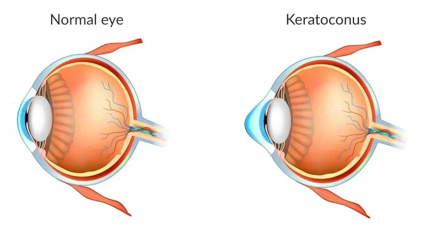
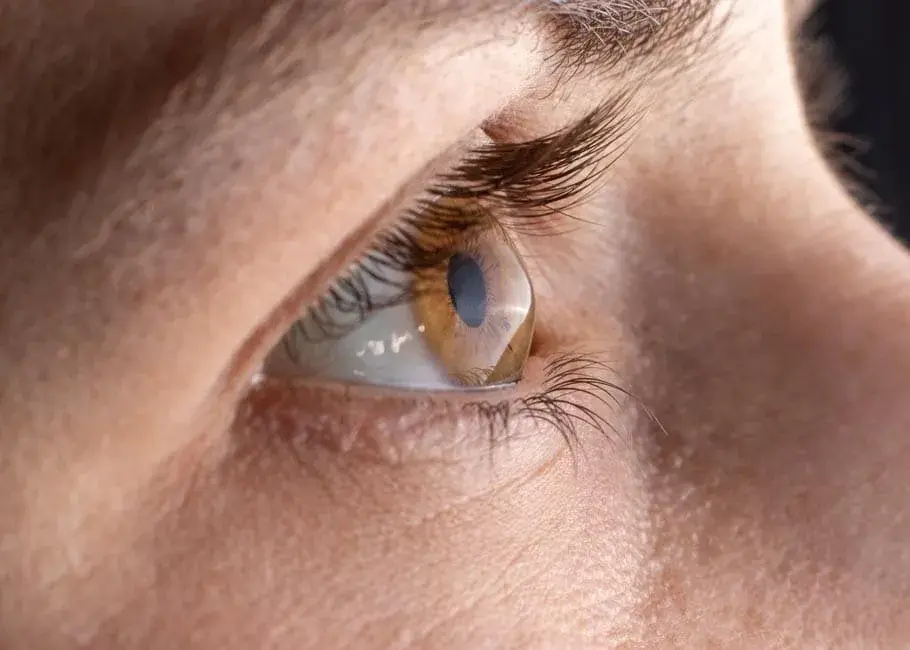
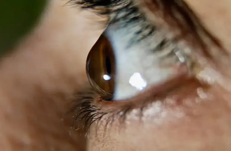
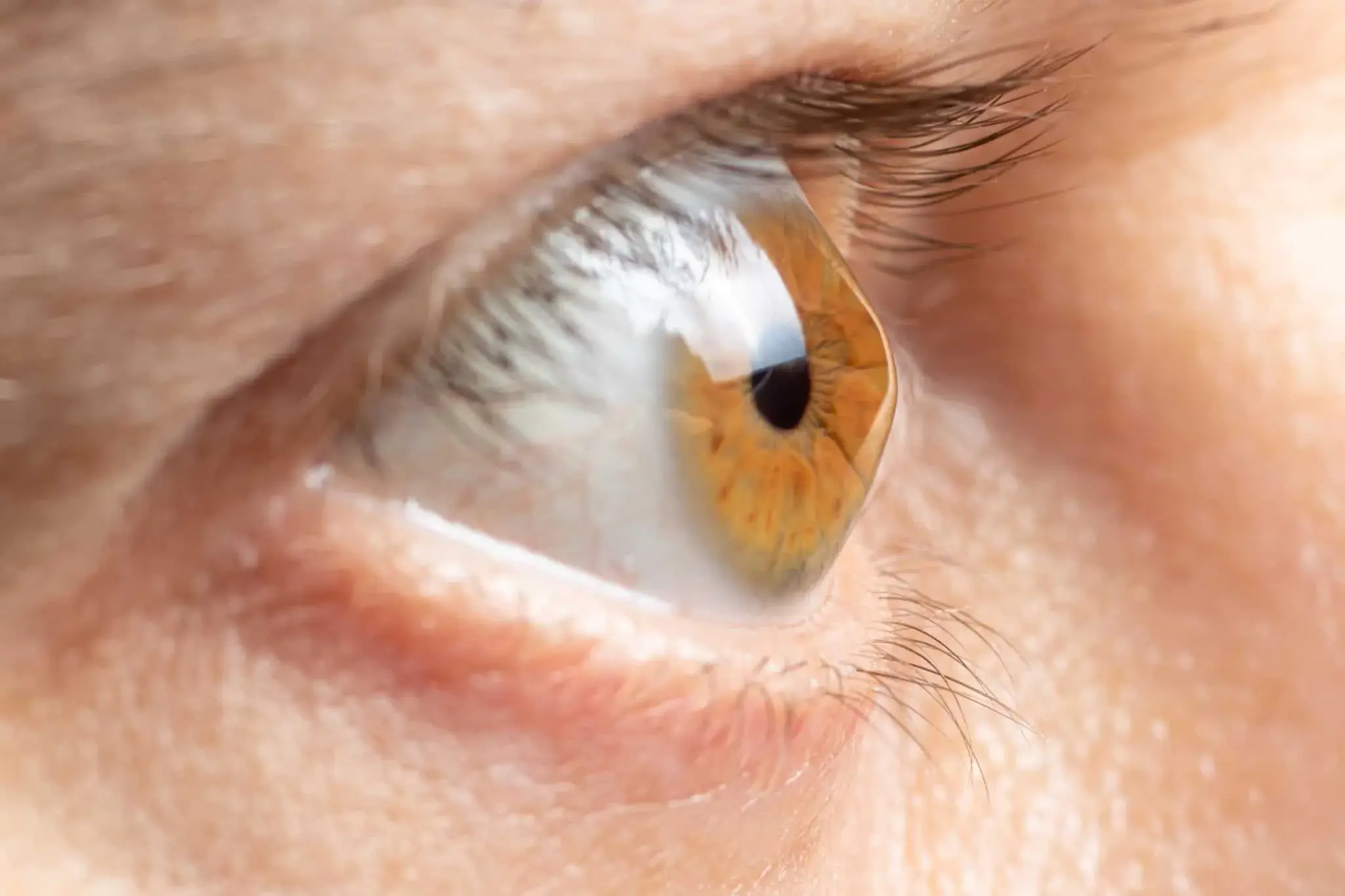
Keratoconus is a progressive eye disorder in which the cornea, the clear, dome-shaped surface at the front of the eye, gradually thins and bulges outward into a cone-like shape. This irregular curvature causes distorted vision, irregular astigmatism, poor-quality night vision, and overall visual disturbances that worsen over time.
In the early stages, many patients manage their condition with corrective lenses, such as glasses or contact lenses. However, as keratoconus advances, these methods may no longer provide adequate vision correction. Patients may begin to experience poor vision that significantly disrupts daily life.
For moderate to severe keratoconus, advanced treatments are often necessary. Options such as corneal cross-linking can stabilize the corneal structure and slow progression. But when the cornea becomes too thin or misshapen, corneal transplantation or other surgical procedures may be considered.
A breakthrough in modern eye care is CAIRS (Corneal Allogenic Intrastromal Ring Segments). This minimally invasive technique reshapes and strengthens the cornea without removing tissue, providing an alternative to traditional corneal transplants. By reinforcing the patient’s cornea and reducing distortion, CAIRS helps many patients achieve improved visual acuity and a better quality of life.
What is CAIRS Eye Surgery?
CAIRS eye surgery represents the next generation of treatments for keratoconus, pellucid marginal degeneration, and post-LASIK ectasia. Performed at the Queensland Eye Institute, the cairs procedure is tailored to the patient’s unique corneal anatomy and designed to stabilize and reshape the eye’s surface.
This surgical procedure involves implanting allogenic tissue ring segments, also known as shaped corneal segments or corneal allogenic intrastromal rings, into the corneal stroma. These cataract segments support and reshape the cornea, helping to improve vision and make patients better candidates for future refractive surgery, such as topography-guided laser resurfacing.
Unlike synthetic Keraring™ implants, CAIRS corneal allogenic intrastromal procedures use donor corneal tissue, which integrates naturally into the host cornea and reduces long-term risks.
Advantages of CAIRS Over Traditional Corneal Transplant
Compared to full corneal transplants or more invasive corneal transplant procedures, CAIRS offers several benefits:
- No corneal tissue removal – CAIRS leaves the patient’s cornea stronger.
- Minimally invasive with minimal pain – Shorter downtime than other surgeries.
- Quick recovery – Most patients return to work within 2–3 days.
- Safe with low complication risks – Less chance of extrusion or rejection.
- Active lifestyle supported – Patients can resume contact sports and swimming within a week.
- Only prescribed eye drops required – Usually just 4 weeks of anti-inflammatory eye drops.
- Reduced long-term risks – No significant chance of graft failure compared with more invasive interventions.
These benefits position CAIRS as a modern solution that delivers excellent visual outcomes while avoiding the complications associated with traditional corneal transplant techniques.
How the CAIRS Procedure is Performed
The cairs surgery process involves several precise steps performed by experienced corneal surgeons:
- Preparation of donor tissue: Tiny allogenic intrastromal ring segments are crafted from donor corneal tissue.
- Laser tunnel creation: A femtosecond laser makes small channels in the patient’s cornea, usually at 50% corneal depth.
- Insertion of corneal ring segments: The donor tissue is inserted into the corneal stroma, reshaping and strengthening the cornea.
- Integration: The intracorneal ring segments become stable within the host cornea, improving light refraction and reducing visual distortion.
Compared to synthetic implants, CAIRS is more effective because it places cairs segments at an optimal depth for reshaping. The modified corneal tissue grafts are non-functioning, meaning even in the rare event of rejection, the corrected visual acuity is not compromised.
Recovery and Vision Improvement After CAIRS
Recovery from CAIRS is usually straightforward and far easier than after full corneal transplants.
- Patients typically need only 2–3 days off work.
- Most patients notice significant improvements in corrected visual acuity within a few days, although their vision may fluctuate as the cornea stabilizes.
- Patients are prescribed anti-inflammatory eye drops and instructed to use them for 4 weeks to reduce inflammation and support healing.
- Follow-up visits ensure proper integration of the cairs segments and to ensure proper healing of the host cornea.
The biggest benefit for many patients is the ability to reduce or sometimes eliminate dependence on corrective lenses. CAIRS can enhance visual acuity, reduce distorted vision, and provide long-lasting improvements in vision.
Who is a Candidate for CAIRS Eye Surgery?
The cairs procedure is particularly suited to patients with:
- Progressive keratoconus not controlled with lenses.
- Moderate keratoconus, where vision is significantly compromised.
- Pellucid marginal degeneration.
- Post LASIK ectasia leading to corneal weakness.
- Patients who are unable to tolerate contact lenses or who have had limited results from corneal cross-linking.
- Individuals seeking an alternative to more invasive interventions like full corneal transplants.
Before surgery, patients undergo detailed eye examinations, including corneal topography, to assess the thickness, curvature, and overall health of their eyes. This information enables surgeons to create a personalized treatment plan tailored to the patient’s unique corneal anatomy.

Insurance Coverage and Costs
While CAIRS surgery is an advanced and highly specialized surgical procedure, it may be covered under private health insurance. In Australia, for example, bronze-level hospital cover or higher often helps cover eye surgery, while extras-only or basic policies typically do not.
For uninsured patients, the CAIRS option can be a significant expense; however, many find the long-term benefits of restored sight and reduced need for corrective lenses to be worth the investment.
Comparing CAIRS with More Invasive Interventions
Unlike traditional corneal transplant methods, CAIRS does not require the removal of the patient’s cornea. Instead, it strengthens the natural tissue with allogenic tissue ring segments, reducing risks while providing excellent visual outcomes.
Full corneal transplants are often reserved for severe cases but involve longer recovery times, higher chances of rejection, and possible graft failure. By contrast, CAIRS is minimally invasive, involves less downtime, and allows patients to return to their everyday activities quickly.
Final Thoughts: Restoring Vision Through CAIRS Eye Surgery
CAIRS eye surgery is a groundbreaking advancement in the management of corneal disorders such as keratoconus. By using donor tissue in the form of intrastromal ring segments CAIRS, surgeons can stabilize and reshape the patient’s cornea, leading to enhanced visual acuity, reduced visual distortion, and lasting vision improvement.
With its minimally invasive approach, shorter recovery, and lower risk of rejection, CAIRS offers hope to many patients who previously faced only more invasive interventions. For those struggling with poor vision caused by progressive keratoconus or related corneal conditions, this procedure represents a safe and effective alternative to full corneal transplants.
As with all corneal procedures, donor corneal tissue plays a vital role. Patients considering CAIRS are encouraged to also discuss organ and tissue donation with their families. The gift of sight can truly transform lives.
