Enhancing Vision with Corneal Allogenic Intrastromal Ring Segments

Corneal allogenic intrastromal ring segments (CAIRS) have emerged as a groundbreaking and minimally invasive treatment for patients with corneal disorders. Designed to reshape the corneal stroma without removing tissue, CAIRS can significantly improve visual acuity and reduce the need for more invasive interventions, such as traditional corneal transplant surgery.
This procedure involves implanting shaped corneal segments made from donor corneal tissue directly into the patient’s cornea. By doing so, surgeons can alter the corneal shape, improve topographic parameters, and enhance overall visual outcomes. Unlike synthetic intrastromal ring segments, allogenic tissue ring segments are biocompatible and better tolerated by the host cornea, reducing the risks of anterior stromal necrosis and other complications.
CAIRS is proving particularly valuable in the management of progressive thinning disorders, such as keratoconus and pellucid marginal degeneration. Under these conditions, the corneal shape becomes distorted, resulting in irregular astigmatism and visual distortion. For many patients, CAIRS not only improves vision but also delays or eliminates the need for corneal transplantation, offering a faster recovery time and a better quality of life.
Conditions Treated with Ring Segments
CAIRS is highly effective in treating various corneal disorders, particularly those involving ectasia or irregular corneal shape. Common indications include:
-
Keratoconus Management – A progressive thinning and bulging of the cornea that leads to distorted vision, irregular astigmatism, and difficulty with night vision.
-
Pellucid Marginal Degeneration – A rarer condition involving thinning of the lower cornea, often causing severe astigmatism and visual distortion.
-
Post-Refractive Surgery Ectasia – Corneal weakening and bulging that can occur after LASIK or other refractive surgeries.
-
Irregular Astigmatism – Complex visual distortions not fully corrected by glasses or contact lenses.
-
Corneal Flattening Needs – Patients requiring shape correction without full corneal transplantation.
By targeting the structural weaknesses in these conditions, CAIRS improves both uncorrected distance visual acuity (UDVA) and corrected visual acuity, helping many patients regain functional vision.
Alternative Vision Correction Methods
While CAIRS is a unique and highly effective treatment, several other vision correction methods exist for patients with corneal disorders. Understanding these alternatives helps patients and ophthalmologists choose the most appropriate approach.
Corneal Cross-Linking (CXL) is often used in conjunction with CAIRS, particularly in the management of keratoconus. It strengthens the cornea by creating additional collagen cross-links, halting disease progression. When combined with CAIRS, CXL provides both structural stability and improved optical performance.
Laser Vision Correction techniques, including LASIK, PRK, and SMILE, reshape the corneal surface to correct refractive errors. However, in cases of thinner corneas or advanced ectasia, laser vision correction may not be advisable, making CAIRS a safer alternative.
Traditional Corneal Transplantation remains the definitive treatment for advanced corneal scarring or severe distortion, but it is more invasive, carries higher complication risks, and requires longer recovery.
Contact Lenses, such as scleral lenses, can mask surface irregularities, but they do not halt disease progression or improve the underlying corneal structure.
The key advantage of CAIRS lies in its ability to preserve the natural corneal structure while offering a customizable and reversible solution with fewer risks than more invasive interventions.

How Corneal Ring Segments Work
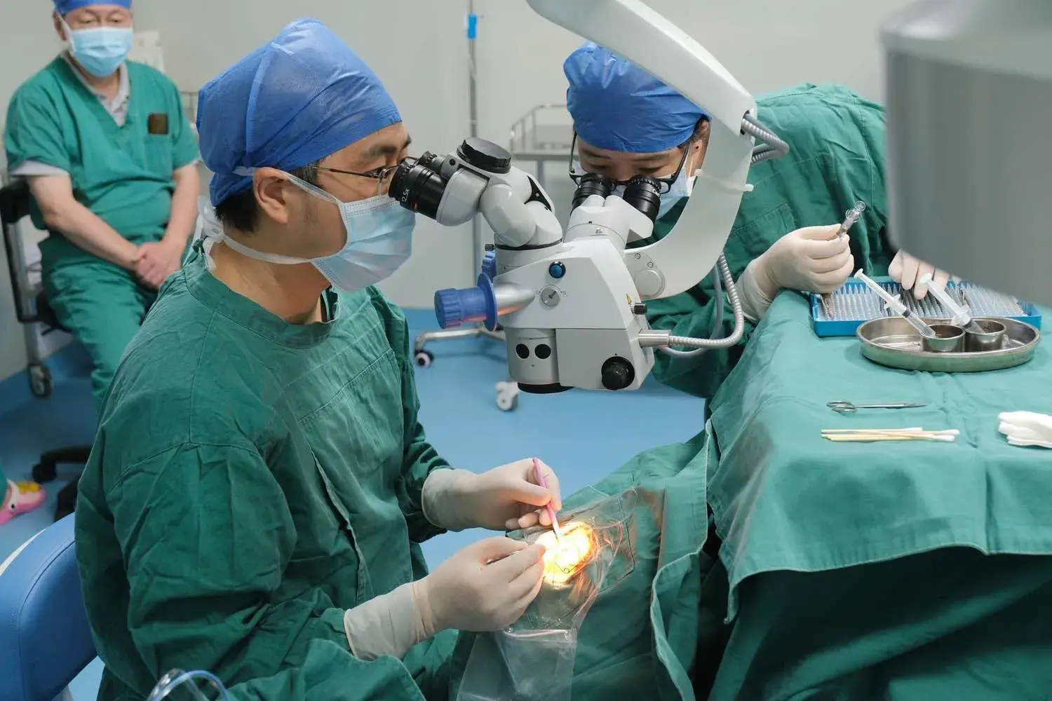
CAIRS uses precision surgical techniques and advanced imaging to reshape the cornea from within. The process begins with a detailed evaluation of the patient’s corneal topography and thickness. This information guides the customization of the intrastromal corneal ring size, inner diameter, and placement based on the patient’s specific needs.
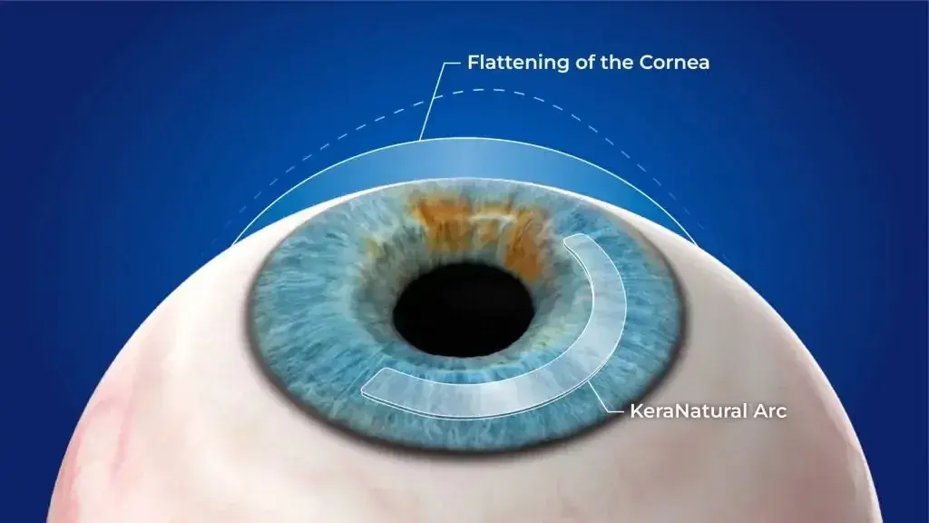
A femtosecond laser creates precise channels in the corneal stroma at predetermined radial clock hours. Into these channels, allograft corneal ring segments—crafted from donor corneal tissue—are carefully inserted. The use of allogenic tissue enhances integration with the host cornea, thereby reducing the immune response and the risk of anterior stromal necrosis commonly associated with synthetic segments.
By adjusting the arc length, thickness, and positioning of the segments, surgeons can reshape the corneal curvature, achieving corneal flattening where needed and improving focus. This tailored approach enables the treatment of decentered asymmetric cones and other irregularities that cause visual distortion.
The technique has a learning curve, but clinical results such as those from the Istanbul Nomogram have shown a significant difference in visual outcomes, patient satisfaction, and long-term stability when compared to synthetic intrastromal ring segments.
Benefits of Allogenic Intrastromal Ring Segments
Patients who undergo CAIRS often experience both optical and lifestyle improvements. Key benefits include:
-
Significant Improvements in Visual Acuity: Better uncorrected and corrected vision in many patients.
-
Reduction in Irregular Astigmatism: Smoother corneal shape for clearer vision.
-
Minimally Invasive Nature: Faster recovery and reduced surgical trauma.
-
Reversibility: Segments can be removed or exchanged if necessary (explantation versus exchange).
-
Lower Risk of Complications: Compared to synthetic segments, allogenic tissue is better tolerated and less prone to causing anterior stromal necrosis.
-
Delay in More Invasive Procedures: Helps avoid or postpone the need for corneal transplantation.
-
Improved Night Vision: Reduced glare and halos for many patients.
These benefits contribute to high patient satisfaction rates, making CAIRS an increasingly preferred option in the management of keratoconus and related conditions.
Candidacy for Vision Correction
The ideal candidates for CAIRS are those with corneal disorders who are not achieving satisfactory vision correction with glasses or contact lenses. Patients with keratoconus, pellucid marginal degeneration, or other forms of corneal ectasia in the early to moderate stages can see substantial improvements.
CAIRS is particularly valuable for patients with thinner corneas, where laser vision correction is contraindicated. It is also an option for individuals experiencing progressive thinning or irregular astigmatism that cannot be effectively managed by non-surgical means.
Before recommending CAIRS, ophthalmologists perform a comprehensive eye examination, including corneal topography, pachymetry, and visual acuity testing. These assessments help determine the optimal intrastromal ring segments CAIRS configuration and evaluate the patient’s overall suitability for the surgical procedure.
Clinical Precision and Customization in CAIRS Surgery
One of the most important factors in achieving successful visual outcomes with CAIRS surgery is the level of precision in planning and execution. CAIRS involves implanting carefully measured CAIRS segments a type of intracorneal ring segments into the corneal stroma to reshape the cornea and improve optical performance.
In the early stages of corneal disorders such as keratoconus, this approach can be particularly effective in halting progression and restoring functional vision. Surgeons often rely on detailed treatment planning methods, including the Istanbul Nomogram clinical results, which provide data-driven guidelines for selecting the appropriate arc length, thickness, and positioning of the ring segments.
This ensures that each procedure is a customized CAIRS intervention, tailored to the patient’s unique corneal topography and visual needs. Published research in Refract Surg journals supports the effectiveness of this individualized approach, showing that careful planning and precise placement of the segments not only enhance visual acuity but also maintain long-term corneal stability.

Procedure and Recovery with CAIRS
Procedure Steps:
-
Performed under topical anesthesia for patient comfort.
-
A femtosecond laser creates channels in the corneal stroma.
-
Customized allogenic tissue ring segments are implanted.
-
Surgery typically takes 20–30 minutes per eye.
Recovery Guidelines:
-
Minimal discomfort post-surgery.
-
Avoid eye rubbing for several weeks.
-
Use prescribed eye drops as directed.
-
Resume normal activities within a few days, avoiding strenuous exercise for the first few days.
-
Attend all scheduled follow-up appointments to monitor the healing process and adjust segments as necessary.

Risks, Costs, and Long-Term Effectiveness
While CAIRS is generally safe, potential complications include infection, delayed healing, and rare cases of donor tissue rejection. These risks are significantly reduced compared to synthetic intrastromal ring segments. Patients should discuss the potential benefits and risks thoroughly with their ophthalmologist before proceeding.
The cost of CAIRS varies depending on the complexity of the case, the number of segments used, and the surgeon’s expertise. Some insurance providers may cover part of the cost, especially when the procedure is deemed medically necessary for conditions like keratoconus. Financing plans are also available in many clinics to make the surgery more accessible.
Long-term studies have shown that most patients maintain their visual improvements for years after surgery. Many experience a sustained reduction in spherical equivalent error and improved corneal topography. These stable outcomes, combined with the procedure’s reversibility, make CAIRS an attractive solution for those seeking lasting vision improvement without undergoing corneal transplantation.
Conclusion
Corneal allogenic intrastromal ring segments are transforming the way ophthalmologists manage corneal disorders such as keratoconus and pellucid marginal degeneration. By using donor corneal tissue to reshape the patient’s cornea, CAIRS offers a biocompatible, minimally invasive, and customizable solution that improves visual outcomes and preserves the natural corneal structure.
For patients struggling with distorted vision, irregular astigmatism, or progressive thinning, CAIRS provides a path toward improved visual acuity and quality of life. With proper patient selection, skilled surgical technique, and ongoing follow-up, the results can be both impressive and long-lasting, making CAIRS a leading choice in modern vision correction strategies.