Corneal Cross Linking Surgery: A Comprehensive Guide to Treating Progressive Keratoconus

Corneal cross-linking surgery is a relatively new treatment designed to strengthen the cornea and prevent the progression of conditions like progressive keratoconus and pellucid marginal degeneration. This outpatient procedure utilizes a combination of ultraviolet light and vitamin B2 eye drops to form new bonds between the collagen fibers in the cornea. Reinforcing the corneal tissue helps maintain a healthier corneal shape, improves visual stability, and reduces the risk of needing a corneal transplant in the future.
In this guide, we will explore how corneal cross-linking works, what to expect before and after surgery, potential risks, and frequently asked questions. Whether you’re in Australia or anywhere else in the world, this article provides a detailed understanding of this treatment.
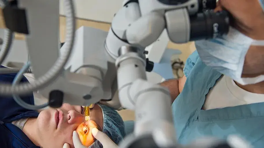
Understanding Corneal Cross-Linking Surgery
Corneal cross-linking surgery (CXL), sometimes called corneal collagen cross-linking or corneal collagen crosslinking, is the only procedure that targets the root cause of corneal weakening rather than just the symptoms.
This treatment is most commonly used for early keratoconus, a progressive condition in which the cornea thins and bulges outward, leading to vision problems such as blurred vision and irregular astigmatism. It can also be performed for other corneal ectatic disorders like progressive corneal ectasia after LASIK or pellucid marginal degeneration.
The aim is to strengthen the collagen fibers in the cornea through a photochemical reaction involving UV light and riboflavin (vitamin B2). The UV light triggers chemical bonds between the collagen molecules, increasing corneal strength and stopping keratoconus progression.
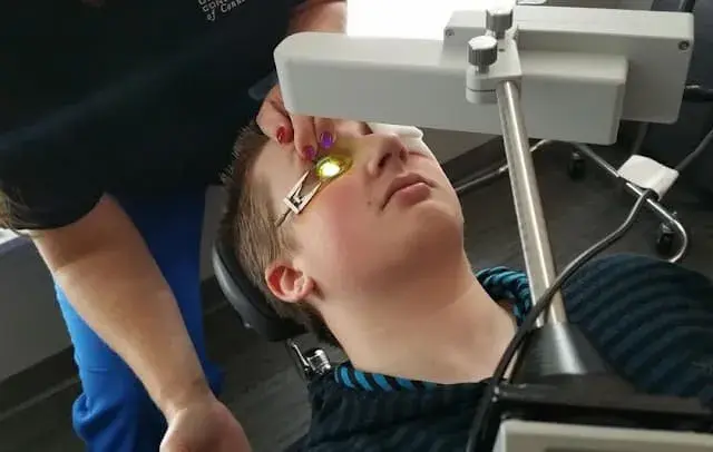
How Corneal Cross-Linking Works
- Preparation and Anaesthesia: The procedure is usually performed under a local anaesthetic, keeping the eyes healthy and pain-free during surgery. In the epi-off technique, the outer layer (epithelium) is removed, while in the epi-on technique, it is left intact.
- Riboflavin Eye Drops: Riboflavin eye drops are applied to saturate the corneal tissue. These drops are crucial for allowing ultraviolet light to penetrate and form new bonds between collagen fibers.
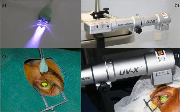
- UV Light Activation: A focused beam of UV light is directed at the cornea for about 30 minutes, initiating a photochemical reaction that strengthens the cornea’s structural framework.
Candidates for Corneal Cross-Linking
Not everyone is suitable for corneal cross-linking surgery. The best candidates are younger patients or those in the early stages of keratoconus. The only treatment proven to halt disease progression, CXL, is particularly beneficial when severe changes in corneal shape have not yet occurred.
Patients with severe corneal scarring may not benefit as much, and in such cases, a corneal transplant might be the better option. For eyes that are healthy enough and have adequate corneal thickness, the results are usually long-lasting.
Corneal topography is essential before deciding on treatment. This imaging maps the corneal shape and detects subtle changes before they cause significant vision problems.
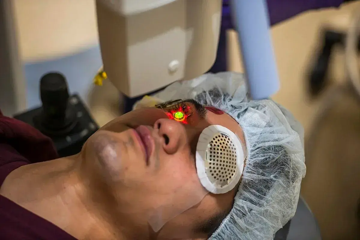
Types of Corneal Cross-Linking
- Epi-Off CXL: Involves removing the outer layer of the cornea for maximum riboflavin absorption. This approach has strong evidence for halting keratoconus progression but requires a longer healing time.
- Epi-On CXL: Maintains the outer layer integrity, resulting in faster recovery but slightly reduced riboflavin penetration.
- Accelerated CXL: Uses a stronger, focused beam of UV light over a shorter duration, reducing the time the procedure takes while aiming for similar effectiveness.
What to Expect During the Corneal Cross-Linking Procedure
The corneal cross-linking procedure is typically performed as an outpatient procedure, lasting between 60 and 90 minutes. After numbing the eye with drops, the surgeon will either remove the epithelium (epi-off) or leave it in place (epi-on). Riboflavin drops are then applied at intervals to allow them to soak into the cornea.
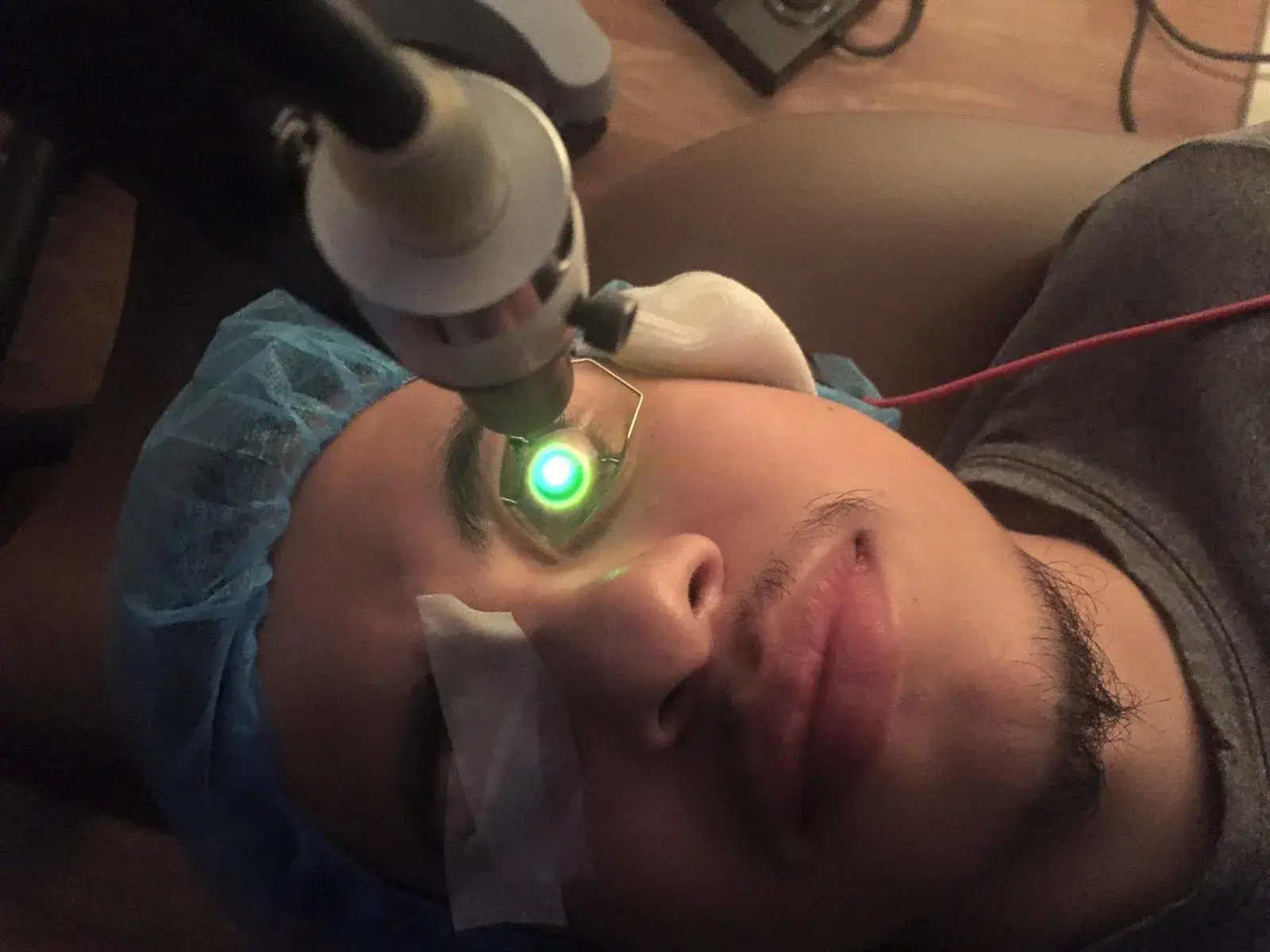
Once saturation is complete, UV light is directed at the cornea for 30 minutes. The photochemical reaction strengthens the collagen fibers by creating new bonds between collagen molecules, thereby stabilizing the cornea.
A bandage contact lens is placed on the eye to promote healing and reduce postoperative pain. You will likely experience some blurred vision and discomfort for about a week, especially with the epi-off method.
Recovery and Follow-Up Care (Bullet Points)
- Immediate Aftercare: Expect to experience blurred vision and mild postoperative pain for the first few days.
- Medication: Steroid drops and antibiotic drops are prescribed to reduce inflammation and prevent infection.
- Follow-Up: A check-up is scheduled within two weeks to monitor healing and assess the best corrected vision improvement.
Risks and Side Effects of Corneal Cross-Linking
While most patients tolerate the procedure well, potential risks include corneal haze, sterile infiltrates, delayed epithelial healing, and temporary light sensitivity. These side effects are more common with the epi-off method but generally resolve over time.
Severe corneal scarring is rare, especially if the surgery is performed on a healthy cornea before advanced disease develops. The treatment is widely considered safe and remains the only procedure proven to halt keratoconus progression in younger patients and those with early keratoconus.
Corneal Cross Linking FAQs
-
How does corneal cross-linking work?
Corneal cross-linking works by applying riboflavin eye drops to the cornea and activating them with UV light. This photochemical reaction strengthens collagen fibres, creating new bonds that increase corneal stability and halt the progression of keratoconus and related corneal conditions.
-
Is it painful?
Thanks to local anaesthetic drops, corneal cross-linking surgery is painless during the procedure. Mild discomfort, irritation, or a gritty sensation is common afterward, especially with the epi-off method, and usually resolves within several days as the epithelium heals naturally.
-
What is corneal crosslinking?
Corneal crosslinking is a minimally invasive procedure that uses UV light and riboflavin to strengthen corneal collagen fibres. It’s an outpatient procedure designed to halt the progression of corneal conditions, making the cornea more stable and preventing further distortion or vision loss.
-
Who should undergo collagen cross-linking?
Patients with progressive keratoconus, pellucid marginal degeneration, or post-LASIK ectasia may benefit from undergoing collagen cross-linking. The procedure is most effective in younger patients or those in the early stages, before significant corneal scarring or thinning limits surgical effectiveness.
-
What is corneal cross-linking (CXL?
Corneal cross-linking (CXL refers to the standardised protocol for strengthening corneal collagen using riboflavin and UV light. It can be performed using epi-off or epi-on techniques and is currently the only treatment proven to halt keratoconus progression in suitable patients.
