
Ectasia After LASIK: Corvis TBI Screening and CAIRS Repair

There are two things I tell every patient who asks me about ectasia after LASIK, and I want to get them down before anything else.
The first is that I don’t do laser on a cornea that can’t take it. Every laser candidate I see has the shape of their cornea mapped with Pentacam tomography and its strength measured with the Oculus Corvis ST, and I combine those two results into a single score called the TBI, which tells me whether the biomechanics are good enough. If they aren’t, we do something else.
The second is that ectasia which has already happened, usually after LASIK done somewhere else, can often be repaired. Other surgeons send me these patients because the cornea can be stabilised with cross-linking and then reshaped with CAIRS keratoplasty, the donor-tissue ring procedure I’ve spent years performing and teaching.
I’ll go through both, starting with what ectasia actually is.
What ectasia after LASIK is
LASIK reshapes the cornea by lifting a thin flap and removing a calculated amount of tissue underneath it with an excimer laser. In the vast majority of eyes the cornea then stays exactly as the laser left it. In a small number, though, it doesn’t. It gradually weakens, thins, steepens and bulges forward, and the vision that was crisp after surgery becomes blurred, doubled and hard to correct with glasses. That’s what we call ectasia. It looks very much like keratoconus, and across large published series it affects roughly 0.04% to 0.6% of eyes treated with LASIK.
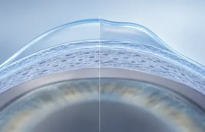
The thing to take from this is that ectasia is a failure of corneal strength. A cornea that develops ectasia after LASIK was almost always weaker than it looked beforehand, and the laser, rather than creating that weakness, took away the margin of tissue that had been covering for it. So when I think about preventing ectasia, I’m really thinking about how well corneal strength was measured before anyone operated.
Why looking at the shape alone isn’t enough
For a long time, screening meant looking at the shape of the cornea. The risk model most people still use was published by Randleman and colleagues in 2008. It scores abnormal topography, how much cornea will be left underneath the flap, age, corneal thickness and the size of the prescription, and of those, abnormal topography is the strongest single warning sign.
Shape-based screening does catch most at-risk corneas, but not all of them, which surprises people. Some eyes that go on to develop ectasia have topography that looks entirely normal before surgery. The weakness is there, it just hasn’t changed the shape yet, and to find those corneas you have to measure the strength itself, because the shape map won’t show it to you.
How I screen: Pentacam tomography plus Corvis biomechanics
Every laser candidate I see has two scans, and each does a different job.
Pentacam tomography maps the front and back surface of the cornea and its thickness at thousands of points. I’ve written before about how tomography changed the way we detect early keratoconus, and this is the shape side of the assessment.
Corvis ST biomechanics looks at strength. The Corvis ST, made by Oculus in Germany, releases a precisely metered puff of air at the cornea and films the cornea bending inward and springing back, using a high-speed Scheimpflug camera running at 4,330 frames per second. From that footage it works out how far the cornea deforms, how fast and how it recovers. A weak cornea bends further and behaves differently from a strong one, even when the two look identical on a shape map, which is the reason I run it.
The TBI score is where the two come together. The Tomographic Biomechanical Index was developed by Renato Ambrósio and colleagues, who used machine learning to combine the Pentacam and Corvis ST data into one number between 0 and 1. In the study that introduced it, the TBI separated ectatic from normal corneas with an area under the curve of 0.996, compared with 0.956 for tomography alone and 0.936 for biomechanics alone. More importantly for LASIK screening, it was built and tested on the hardest cases, the normal-looking eyes of patients whose other eye had ectasia, which are exactly the corneas that shape-based screening misses.
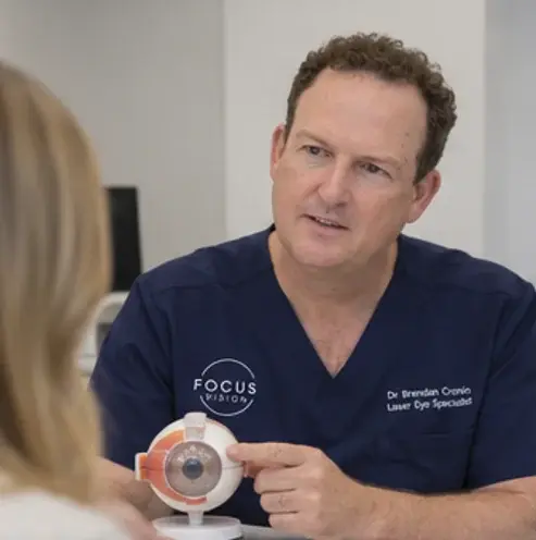
If the TBI and the rest of the assessment tell me a cornea is biomechanically marginal, I won’t do LASIK on it, and I’ll tell you why. That might mean a different procedure, a lens-based option such as an implantable lens or refractive lens exchange, or no surgery at all. I’d far rather turn down a laser procedure than manage an ectasia I caused.
No screening test is perfect, and I don’t pretend this one is. In my view, though, combining shape and strength is the best method we have for making sure a cornea can take the treatment. If you’re considering laser surgery anywhere, ask the surgeon “how have you measured my corneal biomechanics?” If the answer is a topography map on its own, that’s worth knowing before you go ahead.
When ectasia has already happened: repairing it with CAIRS
A good proportion of the post-LASIK ectasia I treat developed after surgery performed somewhere else, and many of those patients are sent to me by their original surgeon or by another ophthalmologist. They refer because the shape of an ectatic cornea can now be rebuilt, and there are two steps to that, done in this order.
First, stop it getting worse
If the scans show the cornea is still changing, corneal cross-linking is how I stabilise it. Riboflavin and ultraviolet light create new bonds between the collagen fibres, which stiffens the cornea. In the US multicentre trial that led to FDA approval of cross-linking for ectasia after refractive surgery, the treated corneas flattened slightly over the first year while the untreated ones kept on steepening. I use epithelium-on cross-linking with the Glaukos Mosaic system, which means a quick recovery for you and a treatment pattern I can customise to the shape of your cornea.
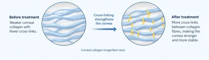
Cross-linking holds the cornea where it is, but it doesn’t reshape it, and that’s where the second step comes in.
Then, rebuild the shape with CAIRS
CAIRS stands for corneal allogenic intrastromal ring segments. They’re arcs of donor corneal tissue that I place into channels cut in the cornea with a femtosecond laser, and they push the bulging part of the cornea back toward a regular curve, which reduces the steepness and the irregular astigmatism that glasses can’t correct. Because the segments are human corneal tissue rather than plastic, they avoid the complications that led synthetic rings such as Intacs to fall out of favour, and CAIRS is performed as a keratoplasty procedure that Medicare recognises. I’ve compared the two in CAIRS vs Intacs and Kerarings.
CAIRS was developed for keratoconus, and that’s still most of what I use it for. Its use in post-LASIK ectasia is newer, and the evidence is growing. A 2025 series in the Journal of Refractive Surgery combined customised CAIRS with cross-linking for post-LASIK ectasia and reported improvement in both uncorrected and best-corrected vision, with reduced corneal steepness and coma and no major complications. That matches what I see in my own patients. Planning the segments for a post-LASIK cornea is a customisation exercise, which is what CAIRSplan, the planning tool I co-developed, is for, and it’s part of what I cover when teaching CAIRS to surgeons internationally.
Once the cornea is stable and more regular, whatever prescription is left gets handled with glasses or contact lenses. For some patients a scleral lens over a reshaped cornea is the combination that gets them back to driving comfortably. If you’d like more, I’ve written about what recovery looks like week by week, how long CAIRS lasts and being asleep for CAIRS surgery.
Will I need a corneal transplant?
Usually not, though it’s often the first thing people ask. Corneal transplantation is still the answer for advanced ectasia with significant scarring, or a cornea too thin for anything else, but it sits at the far end of the pathway. With cross-linking to stabilise the cornea, CAIRS to reshape it and modern lenses to fine-tune the result, most patients never get there.
For referring surgeons and optometrists
If you have a patient with ectasia after LASIK, I’m happy to see them. Send the current tomography, any earlier scans, and the original treatment record if it’s available, since the flap thickness and ablation depth help with planning. Details are on the referrers page.
The bottom line
To come back to where I started, ectasia after LASIK is a strength problem at both ends. Before surgery, my job is to measure corneal strength properly and not laser a weak cornea, which is what the Pentacam, the Corvis and the TBI score are for. If it’s already happened, the job is cross-linking to stabilise the cornea and CAIRS to rebuild its shape. I do both here.
If you’ve noticed worsening vision, increasing astigmatism, ghosting or glare after LASIK, or you’d like your corneal biomechanics measured before you have it, book a consultation.
Frequently Asked Questions
What is the TBI score?
The Tomographic Biomechanical Index combines two scans into one number between 0 and 1: Pentacam tomography, which maps the shape and thickness of the cornea, and Oculus Corvis ST biomechanics, which measures how the cornea bends and recovers when a puff of air hits it. It was developed by Renato Ambrósio and colleagues using machine learning, and in the study that introduced it, it separated ectatic from normal corneas more accurately than either scan on its own.
Does a good TBI score guarantee I won't get ectasia after LASIK?
No test can promise that, and I won't. What the TBI does is measure corneal strength directly rather than inferring it from shape, which is how the corneas that shape-based screening misses get picked up. It is one part of a full assessment that also looks at corneal thickness, the tissue that will remain under the flap, your age and your prescription. Together they are the best method available for deciding whether a cornea can take laser treatment.
What happens if my TBI score is borderline?
I won't do LASIK on a cornea whose biomechanics are marginal, and I'll explain why. Depending on your prescription and your eyes, the alternative may be a different procedure, a lens-based option such as an implantable lens or refractive lens exchange, or no surgery at all. I would much rather decline a laser procedure than treat an ectasia it caused.
I had LASIK elsewhere and now have ectasia. Can it be repaired?
Often, yes. The first step is to stop the cornea changing, which is what corneal cross-linking does. The second is to rebuild its shape, which is what CAIRS keratoplasty does: arcs of donor corneal tissue placed into channels in the cornea that push the bulge back toward a regular curve. Many of the post-LASIK ectasia patients I treat are referred by their original surgeon or another ophthalmologist for exactly this.
Why CAIRS rather than plastic ring segments like Intacs or Kerarings?
CAIRS segments are human corneal tissue, so they avoid the erosion, extrusion and infection problems that led synthetic rings to fall out of favour, and they are performed as a keratoplasty procedure that Medicare recognises, which synthetic rings are not. I have written a full comparison in the CAIRS vs Intacs and Kerarings post.
Will I need a corneal transplant?
Usually not. Corneal transplantation is still the answer for advanced ectasia with scarring or a cornea too thin for anything else, but with cross-linking to stabilise, CAIRS to reshape and modern contact lenses to fine-tune, most patients never get to that point.
